A lo largo de este blog se pueden encontrar los contenidos presentados a los alumnos de Biología y Geología de 3º de ESO y otros cursos
, además de diferentes noticias de contenido científico. Pretende ser un espacio de reflexión y una puerta abierta hacia la observación de nuestro entorno.
They show a remarkable extracellular matrix, which is a scaffolding (andamiaje) made up of collagen and elastic fibers, and other molecules.
The type and proportion of these components in the extracellular matrix set the structural, mechanical and biochemical properties of the different connective tissues.
Extracellular matrix features and cell types define the variety of connective tissues.
Functions
they connect and keep together many organs of the body,
they provide mechanical support to different parts of the body and to the whole body as well,
and protect and isolate many organs.
Furthermore, they allow communication between different parts of the body.
The Connective tissue proper contains several types of cells and a variable amount of extracellular matrix made up of fibers and ground substance (sustancia fundamental)
This tissue is widespread throughout the body. It fills spaces between organs (Figure 2), for example between the skin and muscles, and surrounds blood vessels, nerves and several organs
ADIPOSE TISSUE
It can be regarded as a rather unusual connective tissue since it has very little extracellular matrix.
Adipocytes are the cells that form the adipose tissue, and they have the ability of synthesizing and storing large lipid droplets in the cytoplasm.
These fat depots provide lipids that are used by other tissues to produce energy or just heat
Adipose tissue is not just for lipid storage. It is also involved in the control of the body metabolism by releasing several hormones,
Two types of adipose tissue are found:
white fat (or unilocular) with adipocytes containing a large lipid droplet, and
brown fat (or multilocular) with adipocytes containing many small lipid droplets.
Cartilage is a semi-rigid structure that maintains the shape of several organs, covers the surface of bones in the joints, and is the main supporting tissue during embryonic development, when bones are not yet present. During development, bone substitutes cartilage by endochondral ossification
Ccartilage is mostly an avascular tissue, lacking blood and lymphatic vessels, and without nerve terminals.
Cells that form the cartilage are called chondrocytes. They are located through the tissue in scattered small cavities known as lacunae
BONE
Bones support the soft parts of the body and protect organs like the brain, lungs and heart. They also work as a lever used by muscle to produce movement.
As a metabolic center, bones store calcium and phosphorous, and regulate their metabolism. In addition, in the internal cavities of bones, the bone marrow is made up of many stem cells (hematopoietic cells) that differentiate into blood cells, a process referred as hemaotopoiesis.
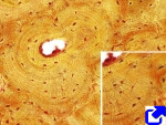
Osteocytes are the most abundant cell type in the mature bone. They are locked in extracellular matrix cavities (bone lacunae
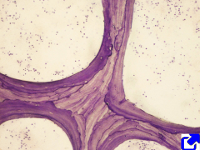
Compact or cortical bone has no vascular cavities, and the extracellular matrix is organized into bone lamellae, which can be arranged straight and parallel (lamellar compact bone) or concentrically around a canal (osteonic compact bone).
Blood is regarded by many authors as a specialized type of connective tissue composed of cells, cell fragments and a liquid extracellular matrix known as blood plasma
Blood has many functions. The following are three salient functions.
1) Communication pathway. Blood transports nutrients and oxygen from intestine and the lungs, respectively, to the rest of the body. Wasting products are transported to the kidney and lungs. It is also the main communication pathway for chemical signals, like hormones, between distant cells in the body.
2) Homeostasis. Blood contributes to the general body homeostasis. For example, it keeps relatively constant the body temperature and pH of tissues.
3) Defense. Blood contributes in the repairing of wounds by sealing the damages with erythrocytes, platelets and plasma, i.e., blood clotting (coagulación). It also contains the cells of the immune system, that use the circulatory system to be transported and attack pathogen in any tissue of the body.
1. Blood cells
Blood cells are classified in two groups: erythrocytes or red cells, and leukocytes or white cells (Figures 1 and 2). There are also cell fragments in the blood referred as platelets.(plaquetas)
Plasma
Plasma is the liquid part of the blood and accounts for more than half of the blood volume. It is 90 % water, and the rest is composed of proteins, ions, amino acids, lipids, and gases. Plasma is the main transporter of nutrients and waste products.
epithelial cells are tightly attached to one another by macromolecular adhesion complexes.
Some epithelia show a high rate of cell turnover where cell death and cell proliferation are frequent.
Some epithelial cells can have apical specializations that allow them to function as sensory receptors, and some animals show complex structures in their epithelial layer, such as hair, feathers or scales.
Epithelia are usually classified according to two features: the number of cell layers and the shape of the cells of the more superficial layer
Simple epithelia are single cell layers where all the cells contact the underlying basal lamina and have an apical free surface. The shape of the cells can be flat (wider than high), cuboidal (as wide as high), or columnar (higher than wide).
Pseudostratified epithelia are simple epithelia where all cells contact the basal lamina, but not all cells reach the superficial layer because some are shorter than the others. Thus, this is a simple epithelium that looks stratified.
Stratified epithelium contains two or more layers of cells. Only cells of the deeper layer are in contact with the basal lamina and only cells of the upper layer show free surfaces.
Stratified epithelia can be classified as
squamous,
cuboidal and
columnar, depending on the shape of the cells of the upper layer when observed in transverse view.
Secretion is the release of substances from cells to the extracellular milieu, having these substances a physiological purpose. Secretory cells are usually grouped to from glands, although it is not always so.
Exocrineglands release their products to the external surfaces of the body (for example, skin, respiratory ducts and digestive duct).
If the products are released into inner tissues and blood stream they are called endocrine glands.
Exocrine glands release their products to internal cavities or to the exterior of the body.
Crenation: A process resulting from osmosis in which red blood cells, in a hypertonic solution, undergo shrinkage and acquire a notched or scalloped surface
Notched: Con muescas
Scalloped: Festoneada
Active transport
Endocytosis: the taking in of matter by a living cell by invagination of its membrane to form a vacuole
Pinocytosis: the ingestion of liquid into a cell by the budding of small vesicles from the cell membrane.
Phagocytosis: the ingestion of bacteria or other material by phagocytes and amoeboid protozoans.
Phagosome: a vacuole in the cytoplasm of a cell, containing a phagocytosed particle enclosed within a part of the cell membrane. Osmosis, the spontaneous passage or diffusion of water or other solvents through a semipermeable membrane (one that blocks the passage of dissolved substances—i.e., solutes
Conective Tissues
Connective tissues have been regarded as a supporting tissue because
they connect and keep together many organs of the body,
they provide mechanical support to different parts of the body and to the whole body as well,
and protect and isolate many organs.
Furthermore, they allow communication between different parts of the body. For example, the molecules absorbed by epithelia must cross the connective tissue before they reach other tissues, so that some connective tissues are considered internal environment of the body.